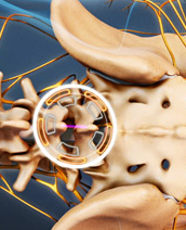
Microdiscectomy
(Minimally Invasive)
In a microdiscectomy or microdecompression spine surgery, a small portion of the bone over the nerve root and/or disc material from under the nerve root is removed to relieve neural impingement and provide more room for the nerve to heal.
A microdiscectomy is typically performed for a herniated lumbar disc and is actually more effective for treating leg pain (also known as radiculopathy) than lower back pain.
Impingement on the nerve root (compression) can cause substantial leg pain. While it may take weeks or months for the nerve root to fully heal and any numbness or weakness to get better, patients normally feel relief from leg pain almost immediately after a microdiscectomy spine surgery.
How Microdiscectomy Surgery Is Performed
A microdiscectomy is performed through a small (1 inch to 1 1/2 inch) incision in the midline of the low back.
- First, the back muscles (erector spinae) are lifted off the bony arch (lamina) of the spine. Since these back muscles run vertically, they can be moved out of the way rather than cut.
- The surgeon is then able to enter the spine by removing a membrane over the nerve roots (ligamentum flavum), and uses either operating glasses (loupes) or an operating microscope to visualize the nerve root.
- Microdiscectomy Surgery Video: A Spine Surgeon Explains the Procedure Microdiscectomy Surgery Video
- Often, a small portion of the inside facet joint is removed both to facilitate access to the nerve root and to relieve pressure over the nerve.
- The nerve root is then gently moved to the side and the disc material is removed from under the nerve root.